Fig. 1.
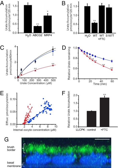
ABCG2 is a urate transporter. (A) C-14 urate accumulation data from Xenopus oocytes injected with either H2O or mRNA coding for MRP4 or ABCG2. (B) Urate accumulation in oocytes injected with H2O, ABCG2 WT mRNA (incubated with or without 5 μM FTC), or the nonfunctional ABCG2 mutant S187T (N = 10 samples each and n = 20 oocytes each for all accumulation experiments). (C) Urate accumulation is dependent on the extracellular urate concentration (H2O [blue circles]; S187T [black squares]; WT [red triangles]; N = 10 and n = 20 each). (D) Urate efflux in oocytes incubated overnight in 500 μM C-14 urate as relative efflux over time (H2O [blue circles]; ABCG2 [red triangles]; N = 5 and n = 50 each). (E) Urate efflux depends on the intracellular urate concentration (H2O [blue circles]: slope = 0.01, N = 55; ABCG2 [red triangles]: slope = 0.03, N = 55; P < 0.001). (F) FTC (5 μM) inhibits urate export in native LLC-PK1 renal proximal tubule cells as shown by increased urate accumulation in FTC-treated cells compared to non-treated control cells (N = 6). (G) LLC-PK1 cells express endogenous ABCG2 at the apical brush border membrane. Merged Z sections of LLC-PK1 cells stained with BCRP1 antibody (green) and nuclear DAPI stain (blue; scale bar, 5 μm). Mean ± SEM; **, P < 0.001; *, P < 0.01.

