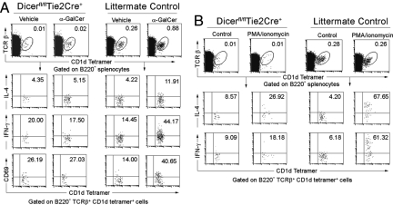
Fig. 5.
Impaired iNKT cell function upon in vivo α-GalCer treatment and in vitro PMA/ionomycin stimulation. (A) IL-4 and IFN-γ production by splenic iNKT cells was analyzed after α-GalCer or vehicle injection by intracellular cytokine staining. Events shown are gated on B220-negative, TCRβ-positive, CD1d tetramer-positive events. α-GalCer or vehicle-stimulated iNKT cells were also analyzed for the expression of the activation marker CD69. (B) Whole splenocytes of Dicerfl/flTie2cre+ and littermate control mice were treated with or without PMA and ionomycin for 3 h in vitro. The production of IL-4 and INF-γ by splenic iNKT cells were analyzed with intracellular cytokine staining. Data are representative of 3 experiments.

