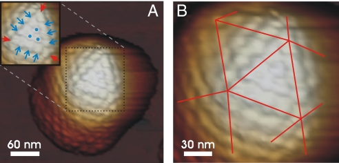
Fig. 3.
AFM images revealing the capsomere organization of icosahedral HSV1 capsids. (A) Image of a B capsid with one triangular face highlighted in the inset. The closed (red) arrows point at the pentons and the open (blue) arrows point at the hexons on the 2-fold axes. The dots mark the 3 central hexons in the middle of the triangular face. (B) Zoom-in on the same particle. The lines mark the borders of the triangular faces of the icosahedral capsid.

