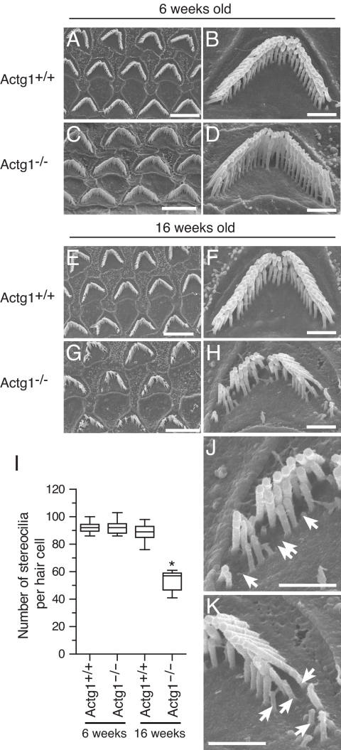
Fig. 4.
Morphology of stereocilia bundles in adult wild-type (Actg+/+) and γcyto-actin deficient (Actg1−/−) mice. (A–D) Scanning electron micrographs of stereocilia from (A, B) 6-week-old Actg1+/+ and (C, D) 6-week-old Actg1−/− mice. (E–H) Scanning electron microscopy images of OHC stereocilia from 16-week-old Actg1+/+ (E, F) and 16-week-old Actg1−/− mice (G, H). There is a loss of individual stereocilia from all three rows of OHC hair bundle from Actg1−/− mice. Images are from the middle turn of the cochlea. (I) Box and whisker plot (whiskers, maximum and minimum; box, 5th–95th percentile; line, mean) of the number of individual stereocilia in individual OHC bundles from Actg1+/+ or Actg1−/− mice at 6 and 16 weeks of age, *P < 0.005. (J–K) enlargements of image in (H) with arrows indicating missing and shortened stereocilia. Scale bars (A, C, E, and G), 5 μm; scale bars (B, D, F, H, J, and K), 1 μm.

