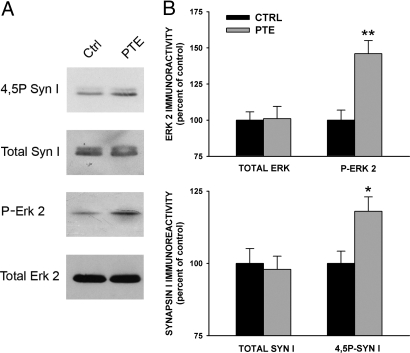
Fig. 4.
Effects of PTE induction in the CA3 area of the hippocampus on the endogenous phosphorylation of ERK 2 and Syn I. Hippocampal slices incubated under basal conditions (Ctrl) or subjected to HFS (50 Hz) to induce PTE were harvested and homogenized. After homogenization, equal amounts of protein (10 μg protein/sample) were subjected to immunoblotting for the total and phosphorylated forms of ERK2 and Syn I, respectively. A representative immunoblot is reported in A. (B) For each slice (n = 5 per experimental group), the amounts of total and phosphorylated ERK 2 and Syn I were quantified and normalized by an internal standard of brain homogenate. Immunoreactivity levels for both total and phosphorylated proteins detected in HFS-treated slices are expressed in percent of the respective levels in control slices (means ± SEM). *, P < 0.05; **, P < 0.01; 1-way ANOVA vs. respective control.

