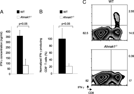
Fig. 4.
In vivo and in vitro deficient IFN-γ production in the absence of AHNAK1. (A) Wild-type and Ahnak1−/− CTLs were obtained as in Fig. S1A, were washed, and were restimulated using plate-bound anti-CD3 only (2 μg/mL) for 24 h. Supernatants from in vitro-stimulated CD8+ T cells were collected, and IFN-γ production was measured by ELISA. Results shown are mean (SD) for 3 mice per group. There is a statistically significant difference between wild-type and Ahnak1−/− cells (P < 0.05). We obtained similar results with 2 other similar experiments. (B) Wild-type dendritic cells pulsed with H-2Kb-binding SIINFEKL peptide (OVA) were transferred into wild-type and Ahnak1−/− mice. Splenocytes were purified and restimulated in vitro with OVA for 6 h, and 7 days later CD8 and IFN-γ expression was analyzed by flow cytometry. The normalized percentages of IFN-γ-producing CD8+ T cells are shown. Results shown are mean (SD) for 6 mice per group. We performed 2 independent experiments. There is a statistically significant difference between wild-type and Ahnak1−/− cells (*P < 0.05). (C) Results from one representative wild-type and Ahnak1−/− are shown.

