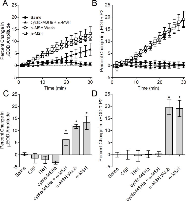
Figure 4.
Effects of peptide and peptidergic factors on the μEOD in vitro. Symbols or bars represent means and error bars show SEM. A) Time course of μEOD amplitude changes in single electrocytes. Alpha–MSH caused rapid μEOD amplitude increases. Cells pretreated with cyclic-MSHa showed a delayed and attenuated μEOD amplitude response in pretreated cells. Following washout of cyclic-MSHa, application of α–MSH rapidly increased μEOD amplitude. B) Time course of μEOD τP2 changes. Alpha–MSH caused rapid and large increases in μEOD τP2. Cells pretreated with cyclic-MSHa showed no change in τP2 in response to α–MSH application. Following washout of cyclic-MSHa, application of α–MSH increases μEOD amplitude and τP2 in a manner indistinguishable from cells that were not pretreated with cyclic-MSHa. C) Percent change in μEOD amplitude after 30 minutes exposure to experimental conditions. Responses to TRH, CRF and cyclic-MSHa were indistinguishable from saline controls after 30 minutes. In contrast, α–MSH caused a large increase in μEOD amplitude. Cells pretreated with cyclic-MSHa showed no change in τP2 in response to α–MSH application, whereas α–MSH produced an attenuated μEOD amplitude response in pretreated cells. Following washout of cyclic-MSHa, application of α–MSH increased μEOD amplitude. D) Percent change in μEOD τP2 after 30 minutes exposure to experimental conditions. Responses to TRH, CRF and cyclic-MSHa were indistinguishable from saline controls after 30 minutes. A–MSH caused a large increase in μEOD τP2, whereas cells pretreated with cyclic-MSHa showed no change in τP2 in response to α–MSH application. Following washout of cyclic-MSHa, application of α–MSH increased μEOD τP2.

