
Figure 2.
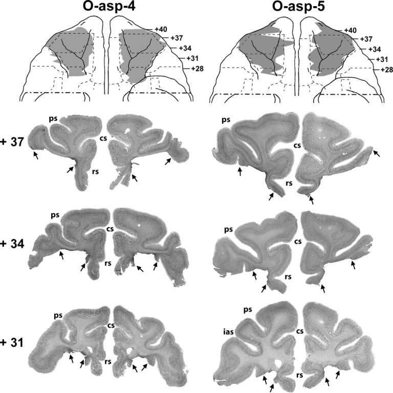
Ventral views of the orbital frontal surface of the monkey brain (top row) depicting the extent of cortical damage in two cases with aspiration lesions (cases O-asp-4 and -5) as estimated from histological sections. Below are photomicrographs of histological sections through the orbital frontal cortex at three anterior–posterior levels. Arrows point to the extent of the lesion at each level. Conventions are as in Figure 1.