
Figure 6.
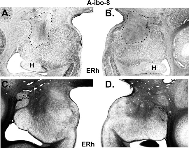
Histological coronal section through the amygdala of case A-ibo-8 that sustained the smallest lesion. A and B display the extent of cell loss as revealed by thionin stain and highlighted with a dashed line. C and D display sparing of fibers (darker areas) as revealed by Gallyas stain in areas where cell loss was almost complete. ERh, Entorhinal cortex; H, hippocampus.