Figure 3.
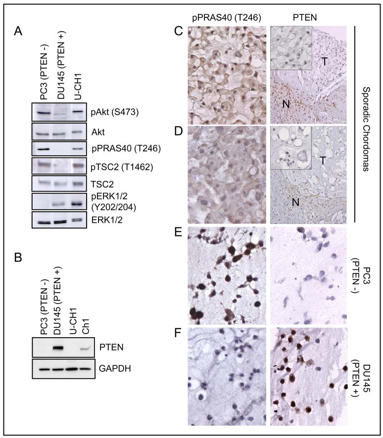
Constitutively activated Akt signaling and PTEN deficiency in chordoma-derived cell lines and sporadic sacral chordoma tumors. A, constitutive phosphorylation of Akt, TSC2, PRAS40, and ERK in U-CH1 cells. Cells were serum-starved for 20 hr before lysates were prepared for western analysis. B, PTEN expression was not observed in U-CH1 cells and significantly reduced in Ch1 cells. PTEN-negative PC3 and PTEN-positive DU145 cells were used as negative and positive controls for PTEN expression. C and D, representative images of two sporadic chordomas showing negative staining with anti-PTEN and positive staining with anti-pPRAS40 (T246) antibodies. N, non-neoplastic normal cells; T, tumor. Magnification: X 400 (left column and insets in right column); X 200 (right column). E and F, PTEN-negative PC3 cells (E) and PTEN-positive DU145 (F) cells showing pPRAS40 and PTEN staining. Magnification: X 400.

