
Figure 1. Intravenously administered reovirus can reduce lymph node metastatic melanoma burden, prime anti-tumor immunity and induce pro-inflammatory cytokines.
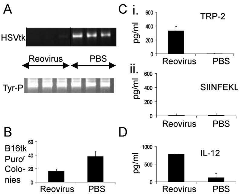
C57Bl/6 mice were seeded with sc B16-tk tumors (5×10⁵ cells). 10 days later, mice were treated intravenously with 5×10⁸ pfu reovirus or with PBS. 10 days after that, tumor draining LN and spleen were explanted, and LN were dissociated and plated overnight in culture. A. Total genomic DNA from 106 LN cells was screened with primers specific for the HSVtk gene. Equal loading of DNA was confirmed using primers specific for a genomic fragment in the tyrosinase gene promoter. B. 106 cells from the dissociated LN cultures were seeded in medium containing 1.25μg/ml puromycin to select for viable B16-tk cells which were present in the LN at the time of resection. Within 5-10 days individual puror colonies appear, which were counted. C. Spenocytes recovered at day 10 were pulsed, in triplicates of 750000, with the synthetic TRP-2 180-188 SVYDFFVWL peptide (i) or with the irrelevant H-2Kb-restricted Ova SIINFEKL peptide (ii). 48h later supernatants were assayed by ELISA for IFNγ. D. Explanted tumor draining LN were dissociated and plated overnight in culture. Supernatants were assayed for IL-12. Data shown representative of one of two independent experiments.