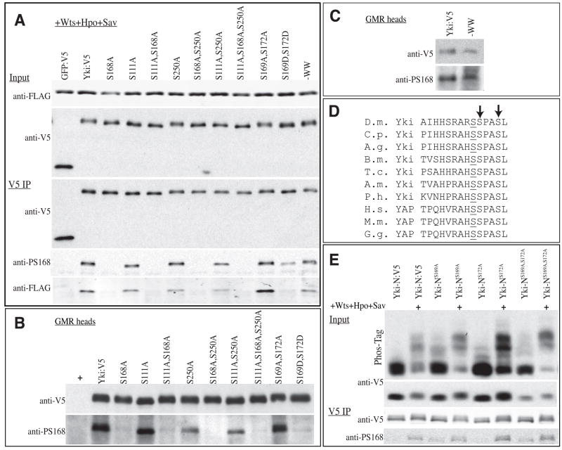
Figure 6. Influence of other phosphorylation sites on Ser168 phosphorylation.
A) Western blots (4–15% gels) of samples from S2 cells co-transfected to express 14-3-3ε:FLAG, 14-3-3ζ:FLAG, Wts, Hpo, Sav, and GFP:V5 or Yki:V5 isoforms. Top two panels show (Input) show blots on cell lysate, using anti-FLAG (14-3-3) or anti-V5 (GFP or Yki) and bottom three panels (V5 IP) show blots on material precipitated by anti-V5 beads. 14-3-3ε andζ have similar mobilities and were not separated. (B,C) Western blots on lysates of adult heads from animals expressing UAS-yki:V5 isoforms under GMR-Gal4 control. Top and bottom panels show the same membrane, blotted with anti-V5 and anti-Yki-S168P, respectively. (D) Amino acid sequence alignment illustrating conservation of Ser residues at +1 and +4 positions (arrows) relative to Yki-Ser168. D.m.: Drosophila melanogaster, C.p.: Culex pipiens (mosquito), A.g.: Aedes aegypti (mosquito), B.m.: Bombyx mori (silkworm), T.c.: Tribolium castaneum (flour beetle), A.m.: Apis mellifera (honey bee), P.h.: Pediculus Humanus corporis (human body louse), H.s.: Homo sapiens, M.m.: Mus musculus, G.g.: Gallus gallus (chicken). (E) Western blots of samples from S2 cells co-transfected to express Yki-N (N-terminal 240 amino acids of Yki) isoforms, and, where indicated, Wts, Hpo, and Sav. Top two panels (Input) show blots on cell lysates, using anti-V5 (Yki), uppermost panel is a Phos-tag gel (25 μM Phos-tag) and below is a 4–15 % SDS-PAGE gel. Bottom two panels (V5 IP) show blots on material precipitated by anti-V5 beads, and run on 4–15% gels, using anti-V5 or anti-Yki-S168P, as indicated.

