
Figure 1. Modulation of H3K9 methylation by Cr(VI).
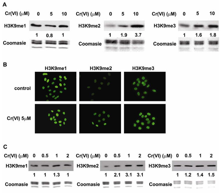
A549 cells were exposed to 5 or 10 μM of Cr(VI) for 1 hour and then the mono- (H3K9me1), di- (H3K9me2), and tri-(H3K9me3) methylation of H3K9 were studied by Western Blot (A) or immunofluorescent staining (B). (C) Beas-2B cells were exposed to 0.5, 1, and 2 μM of Cr(VI) for 1 hour, and the levels of H3K9 methylation were measured by Western Blot. The relative intensity of the bands was measured and is shown below the western blots in the figure. Coomassie blue staining was used to assess the equal loading of histones in Western blot analysis.