
Figure 6. The role of ascorbate in Cr(VI) induced H3K9 dimethylation.
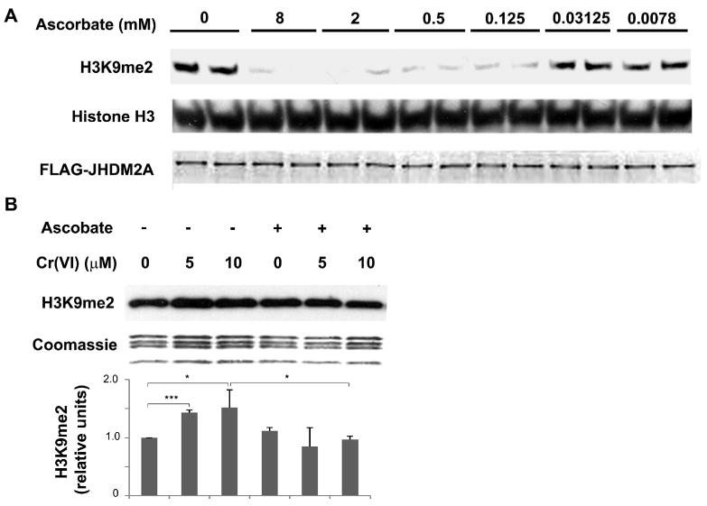
(A) JHDM2A demethylated H3K9me2 in an ascorbate-dependent manner. Purified recombinant Flag-JHDM2A was incubated with core histones in a buffer containing 100 μM Fe3+, 1 mM 2-oxoglutarate, and increased concentrations of ascorbate as indicated. After reaction, the loss of H3K9me2 in histones was assessed using Western blot. The same membrane was blotted with anti-histone H3 antibody to assess the loading of histones. The bottom figure shows the amount of recombinant Flag-JHDM2A added into each reaction. (B) A549 cells were pretreated with 1 mM of ascorbate for 2 hr. After washing with PBS, the cells were exposed to various concentration of chromate for 1 hr. The levels of di-methylated H3K9 were analyzed using Western blot. Coomassie blue staining was used to assess the loading of the histones. The relative intensity of the bands was measured and plotted as means ± SE (error bars). * P < 0.05, ** P < 0.01, *** P < 0.001.