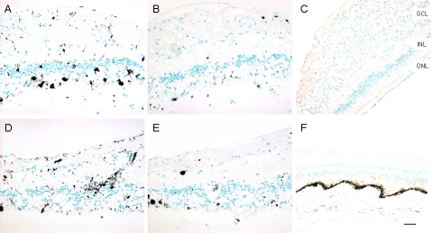
Fig. (5).
Imunoreactivities against CD68, CD3, and CD20 markers in the retinas. A, CD68, right eye; B, CD3, right eye; C, CD20, right eye; D, CD68, left eye; E, CD3, left eye; F, CD20, left eye. CD68 positive cells are scattered in the retina, while most are where photoreceptor are lost. Rare, scattered CD3 positive cells are present in the retina. CD20 staining is negative in both eyes (Green = methyl green counterstain; brown = positive immunostain). (GCL, ganglion cell layer; INL, inner nuclear layer; ONL, outer nuclear layer; Scale bar indicates 50 µm).

