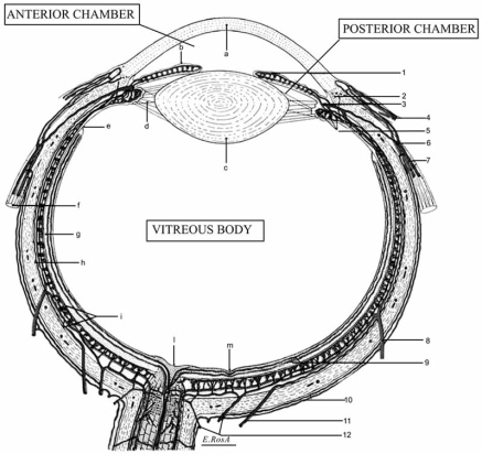
Fig. (3).
Schematic horizontal section of the human eye: anatomic structures and vasculature.
Legend: a, cornea; b, iris; c, lens; d; suspensory ligament of the lens (ciliary zonule of Zinn); e, ora serrata; f, medial rectus muscle of the eye; g, retina; h, sclera; i, choroid; l, optic nerve head; m, central fovea of the macula; n, lamina cribrosa; o, irido-corneal angle.
1, minor circle of iris; 2, Schlemm's channel; 3, major circle of iris; 4, iuxtalimbic conjunctival vessels; ciliary circle; 6, anterior ciliary artery; 7, anterior ciliary arteries and veins; 8, vorticose vein; 9, retinal microvasculature; 10, episcleral arteries and veins; 11, long posterior ciliary artery; 12, short posterior ciliary arteries; 13, central retinal artery and vein; 14, perioptic nerve arteriolar anastomoses (circle of Haller and Zinn); 15, pial microarterioles.

