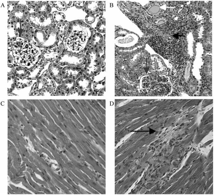
FIGURE 2.
Histologic evaluation of kidneys and hearts. Histologic analysis of kidneys (A, B) and hearts (C, D) from an untreated control (B, D) and from a 9 week CTLA4Ig treated mouse (A, C). Note the presence of vasculitis (horizontal arrow, B) tubular casts (white arrow, B) and glomerulonephritis (vertical arrow, B) in the kidney of the untreated control. Small myocardial infarcts are noted in the control (black arrow) but not in the treated mouse.

