Figure 4.
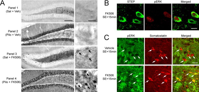
STEP and seizure-induced ERK activation. A, Immunolabeling for the activated form of ERK. Under both the control condition [Saline (Sal) + Vehicle (Veh), Panel 1] and 15 min after SE [Pilocarpine (Pilo) + Veh, Panel 2] onset, ERK activity was not observed in the hilus. In contrast, SE triggered a robust increase in ERK activation in the GCL. FK506 alone (1 mg/kg) triggered ERK activation in the hilus (Sal + FK506, Panel 3). ERK activation was also observed in the hilus after SE in mice given injections of FK506 (Pilo + FK506, Panel 4). The boxed regions are magnified images shown to the right of each panel. Arrows denote cells with marked pERK expression. B, Double labeling for pERK and STEP demonstrates that FK506 injection activated ERK signaling in two of the three STEP-positive hilar interneurons. Representative images were taken from animals killed 15 min after SE onset. C, Double labeling demonstrates that FK506 triggered the expression of pERK in somatostatin-positive cells of the hilus. Animals were killed 15 min after SE onset. Arrows denote somatostatinergic cells. Scale bars: A, 100 μm (low-magnification image) and 25 μm (high-magnification image); B, 10 μm; C, 25 μm.

