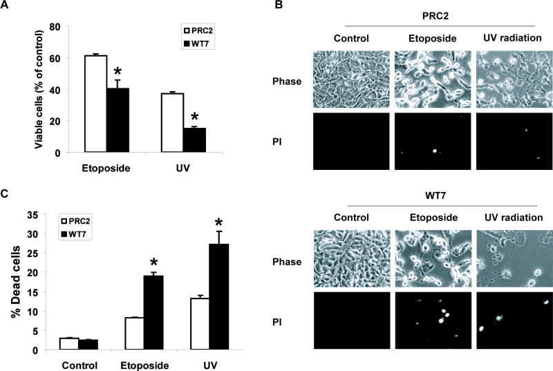
Figure 1. Effect of pVHL expression on the sensitivity of RCC cells to etoposide and UV radiation induced cell death.
(A) Equal numbers of PRC2 and WT7 cells were treated with etoposide (50 μM), exposed to UV radiation (1000 J/m2), or left untreated. After 24 h, the cells were fixed and stained with Hoechst dye in order to visualize nuclear morphology. Viability was assessed by first counting the number of drug or UV-treated cells that retained a normal cellular morphology (viewed under phase-contrast microscopy) and a rounded nucleus containing diffuse Hoechst-stained chromatin. This number was divided by the number of similarly appearing healthy cells counted in the corresponding untreated control, with the quotient converted to a percentage. Data are the mean ± SEM from 3 independent experiments. (B, C) PRC2 and WT7 cells were treated with etoposide or exposed to UV radiation as described above. Twenty-four hours later, the cells were stained with propidium iodide (PI) and digital images were captured from three fields of view in each dish. The percentage of propidium iodide stained cells was determined for each condition with the results representing the mean ± SEM from 3 independent experiments. Exposure to etoposide or UV resulted in a greater reduction in viable cell number and a larger number of dying cells in WT7 cells than in PRC2 cells (*, p<0.05 using ANOVA and Dunnett’s post-hoc test).

