
Figure 4. Pten-deletion-induced invasive adenocarcinoma requires Rictor.
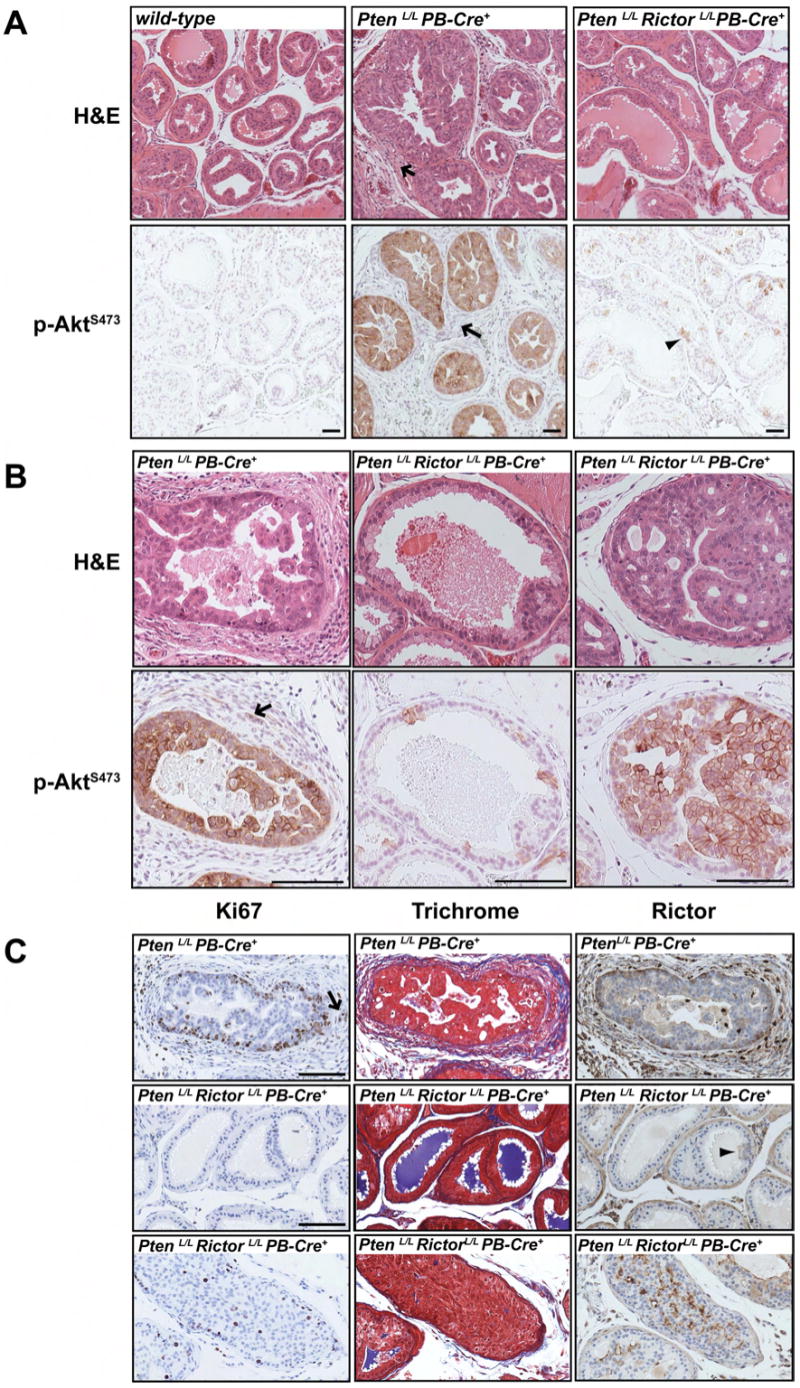
(A) Prostate tissue serial sections from 9-week old mice were stained by H&E or labeled with a phospho-AktS473 antibody and imaged at 10X. The arrows point to changes in the stroma. The arrowhead indicates a patch of phospho-AktS473 positive cells. Scale bar = 50μm. (B) Higher magnification images (20X) of serial sections stained by H&E or labeled with a phospho-AktS473 antibody. Invasive phospho-AktS473 positive cells are indicated with an arrow. Scale bar = 50μM. (C) Labeling with Ki-67 antibody (left), trichrome stain (middle) and Rictor antibody (right) are shown. The arrow indicates proliferating (Ki-67 positive) cells in the stroma. The arrowhead points to a small patch of cells that have not lost Rictor expression. Scale bar = 50μm.