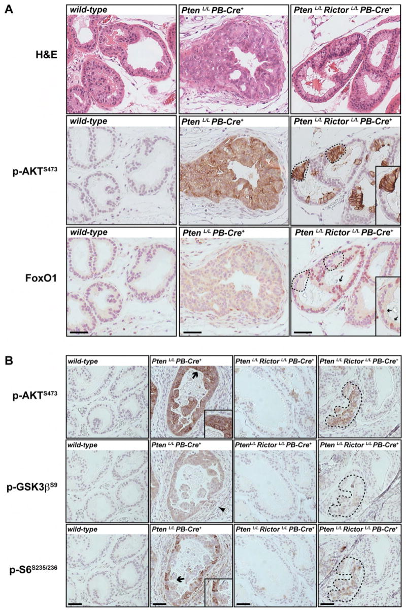
Figure 5. Akt activity towards downstream substrates in Pten-deficient prostate epithelial cells requires Rictor.

(A) Serial sections from wild-type, PtenLoxP/LoxP PB-Cre+ or PtenLoxP/LoxP RictorLoxP/LoxP PB-Cre+ tissue stained by H&E, or labeled with antibodies to phospho-AktS473 or to FoxO1 are shown. The dotted circles indicate phospho-AktS473 positive cells in which Foxo1 is excluded from the nucleus. Arrows point to phospho-AktS473 negative cells in which Foxo1 concentrates in the nucleus. An enlarged section is show in the boxed insert. Scale bar = 25μm. (B) Serial sections from wild-type, PtenLoxP/LoxP PB-Cre+ or PtenLoxP/LoxP RictorLoxP/LoxP PB-Cre+ tissue labeled with antibodies to phospho-AktS473, phospho-GSK3βS9, or phospho-S6S235/236 are shown. Arrows indicate regions highlighted in the boxed inserts. The arrowhead points to invasive cells. The encircled area in the right panels indicates a patch of phospho-AktS473 positive cells. Scale bar = 25μm.
