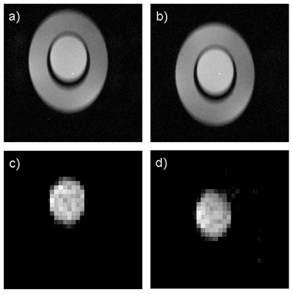
Figure 5.

The phantom structure is clearly depicted in the gradient echo, T1 weighted scout images (a, b). Lactate images generated with the HS-SelMQC-CSI protocol exhibit strong signal from the lactate solution in the inner cylinder with no signal from the lipid in the outer annulus (c, d).
