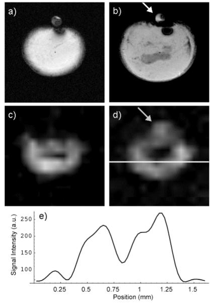
Figure 7.

T1 weighted gradient echo images of a tumor xenograft had a hypo-intense core surrounded by homogeneous tissue (a, b). The lactate signal was suppressed in the tumor core (c, d) and the region of this suppression was larger than the hypo-intense region observed in the gradient echo images. The lactate reference phantom was evident in only one of the two slices in both the gradient echo (b) and lactate (d) images (arrows). A profile of the lactate image along the white line in (d) shows the SNR of the lactate images (e).
