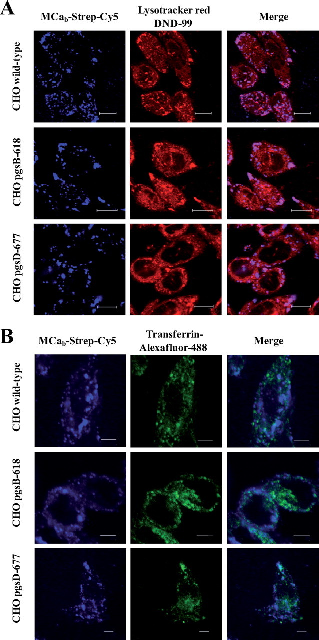
FIGURE 6.

MCab-Strep-Cy5 entry and endocytosis. A, endocytic route of entry of MCab-Strep-Cy5. Various CHO cell lines (upper panels, wild type; middle panels, pgsB-618; lower panels, pgsD-677,) were incubated 2 h with 1 μm of MCab-Strep-Cy5, washed, and incubated with 50 nm LysoTracker red DND-99 for 20 min right before confocal acquisition. Scale bars, 10 μm (upper panels) and 11 μm (middle and lower panels). B, different endocytic entry pathways for transferrin-Alexa Fluor 488 and MCab-Strep-Cy5. Confocal immunofluorescence images of living wild-type or mutant CHO cells to compare the cell distribution of transferrin-Alexa Fluor 488 and MCab-Strep-Cy5. Cells were incubated 2 h with 1 μm MCab-Strep-Cy5 (blue) along with 25 μg/ml transferrin-Alexa Fluor 488 (green), washed, and immediately analyzed by confocal microscopy. Scale bars, 5 μm.
