
Figure 2.
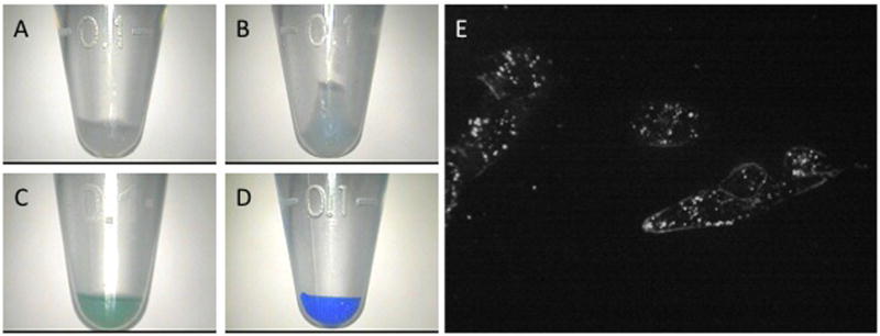
Visible and microscopic tagging of glioma cell pellets by dye-loaded NPs. Cell samples shown have been treated with 1 mg/mL blank NPs (A), methylene blue-loaded NPs (B), indocyanine green-loaded NPs (C), or CB-loaded NPs (D). Duration of treatment is 15 min for samples C and D and 6 h for A and B. Confocal microscopic images of cells treated with 0.0625 mg F3-targeted methylene blue/CB-loaded NPs for 15 min (E).