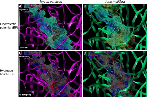
Fig. 8.
Electrostatic (A and B) and hydrogen bonding (C and D) fields extended from the ligand binding site of the peach potato aphid M. persicae (A and C) and the honeybee A. mellifera (B and D). Models were constructed according to Toshima et al. (2009). In A and B, regions with high (positive) and low (negative) electrostatic potentials are colored red and blue, respectively. In C and D, the hydrogen-bond-accepting atoms such as nitrogens and oxygens are colored blue, whereas the hydrogen atoms attached to these hetero-atoms are colored red. In all, carbon, hydrogen, nitrogen, oxygen, and chlorine atoms of imidacloprid are colored white, light blue, blue, red, and blue-green, respectively. The grooves extending from the NO2 binding site in the pest and beneficial species nAChRs differ in terms of the size, electrostatic, and hydrogen bonding properties, which may lead to a generation of pest target-selective insecticides.

