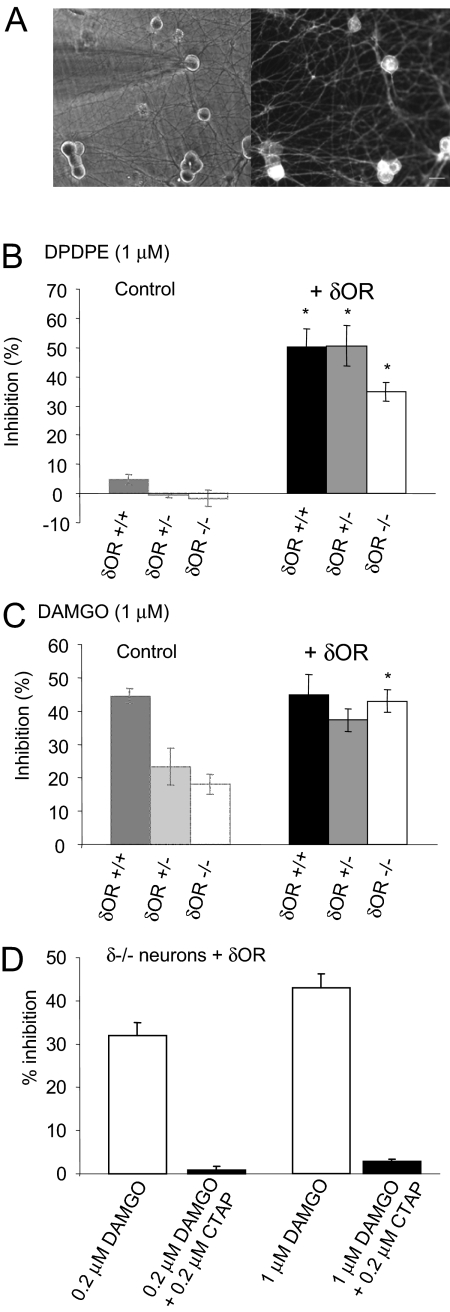
Fig. 5.
Expression of recombinant δ receptors in δ-receptor-deficient neurons restores full μ-receptor coupling. A, Left, electrode, viewed using transmitted light, attached to a cultured DRG neuron. Right, the same neurons viewed using fluorescence microscopy. Labeled neurons contain recombinant δ receptor fused to CFP. B, the bar graph depicts the mean Ca2+ current inhibitions evoked by DPDPE (1 μM) in δ(+/+), δ(+/-), and δ(-/-) DRG neurons expressing recombinant δ receptor (+δOR). Bars on the left provide control data (in the absence of δOR) for comparison (from Fig. 1B). Expression of recombinant δ receptors in neurons of all three genotypes caused a significant increase in the inhibition of VDCCs by DPDPE, *, p < 0.001, Student's t test. C, the bar graph illustrates the mean Ca2+ current inhibitions evoked by DAMGO (1 μM) in neurons expressing recombinant δ receptors. Bars on the left provide the relevant control data for comparison (from Fig. 1B). Expression of recombinant δ receptors in δ(-/-) neurons caused a significant increase in the inhibition of VDCCs by DAMGO, *, p < 0.001, Student's t test. D, the bar graph shows the average inhibition of Ca2+ currents by DAMGO applied to neurons in the presence or absence of the μ-receptor-selective antagonist CTAP. The inhibition of Ca2+ currents by DAMGO, recorded from δ(-/-) neurons expressing recombinant δ receptors, was attenuated by coapplication with the CTAP. In all graphs, the vertical lines represent ± S.E.M.

