Fig. 6.
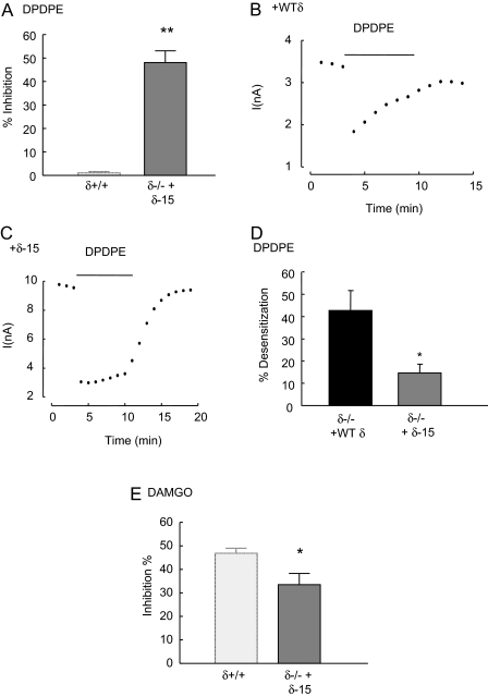
Expression of a heterodimerization-defective δ receptor construct in δ(-/-) neurons does not fully restore μ-receptor inhibitory coupling. A, the bar graph illustrates the mean inhibition by DPDPE (1 μM) of Ca2+ currents recorded from δ(-/-) DRG neurons expressing the dimerization-deficient δ-15 construct that lacks the last 15 amino acids of the δ receptor. The left bar represents the mean inhibition of Ca2+ currents when DPDPE was applied to δ(+/+) neurons (data from Fig. 1B) and is included here for comparison. Like recombinant full-length δ receptors (Fig. 5B), δ-15 receptors mediate robust inhibitory coupling to VDCCs. The dot plots illustrate the time course of the inhibition of Ca2+ currents by DPDPE (1 μM) in δ(-/-) neurons expressing the full-length recombinant δ receptor (+WTδ; B) and the truncated δ-15 construct (+δ-15; C). Note that the full-length δ receptor undergoes desensitization leading to a reduction in the inhibitory effect of DPDPE with time. D, the bar graph depicts the mean percentage of desensitization during 5-min exposure to DPDPE of δ(-/-) neurons expressing the full-length δ receptor or the δ-15 construct. The loss of DPDPE-evoked inhibition after 5 min of exposure was expressed as a percentage of the peak inhibition in each cell. E, the bar graph illustrates the mean inhibition evoked by DAMGO (1 μM) in δ(-/-) neurons expressing the δ-15 receptor. The bar on the left provides for comparison the mean inhibition by DAMGO of Ca2+ currents recorded from δ(+/+) neurons (data from Fig. 1B). The DAMGO-evoked inhibition in cell expressing the δ-15 is less than that of δ(+/+) neurons (p < 0.05) but greater than observed in δ(-/-) (p < 0.05) neurons. In all graphs, the vertical lines represent ± S.E.M. Significance was determined using the Student's t test: **, p < 0.01; *, p < 0.05.

