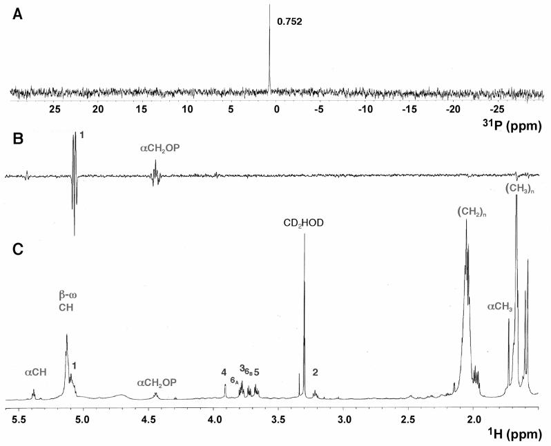
Figure 5.
31P NMR spectrum and selective inverse 31P-decoupled-1H-detected difference spectroscopy of the purified lipid. A. The 31P NMR spectrum of the purified lipid is consistent with the presence of one monophosphodiester (0.752 ppm). B. The selective inverse decoupled difference 1H NMR spectrum obtained with on and off resonance 31P decoupling of the 0.752 ppm phosphorus signal (30, 31) showed that the anomeric carbon of GalN is linked via a phosphodiester bridge to the proximal isoprene unit of the undecaprenyl group. C. The one-dimensional 800-MHz 1H NMR spectrum of the donor lipid shows six GalN and various isoprene methine, methylene and methyl proton signals (see Tables 1 and 2 for details). Note that an impurity peak overlaps H-5 near 3.66 ppm. The proposed structure of undecaprenyl phosphate-β-d-GalN is shown in Fig. 2 along with the numbering scheme.

