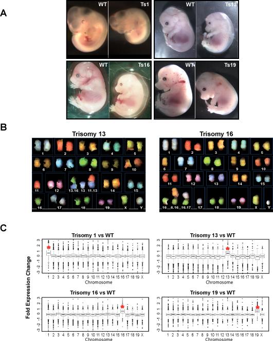
Fig. 1. Generation of trisomic embryos and mouse embryonic fibroblast (MEF) cell lines.
(A) Trisomic (Ts) embryos were recovered by timed matings. The trisomy 1 (Ts1) embryo was recovered at 10.5 days postcoitum (dpc). Ts13, Ts16 and Ts19 embryos were recovered at 14.5 or 15.5dpc. In all instances Ts embryos were identified by their developmental abnormalities and reduced size (7).
(B) Examples of Spectral karyotype (SKY) analysis of metaphase spreads prepared from early passage (≤p3) Chr13 and Chr16 trisomic MEFs. Chromosomes and Robertsonian translocations are identified.
(C) Gene expression pattern of aneuploid cell lines and lines from euploid littermate controls (12). Transcripts were binned by chromosome and the average gene expression/total chromosome is shown. The asterisk indicates the identity of the trisomic chromosome. The increase in gene expression was highly significant (p≤ 1x10−74, all trisomies, Student's t-Test).

