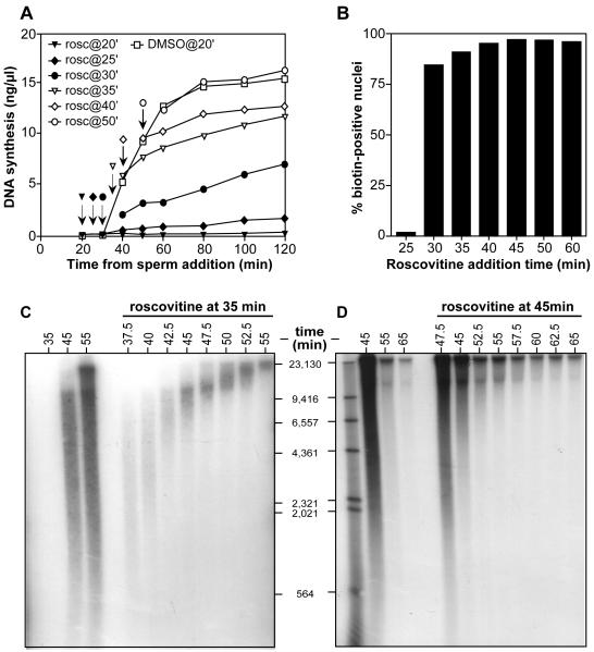
Figure 2.
Inhibition of CDK activity prevents later origins from firing.
(A) Sperm nuclei were incubated at 15 ng DNA/μl in egg extract supplemented with [α-32P]dATP. Aliquots were supplemented with 0.5 mM roscovitine at the following times: filled triangles, 20 min; filled diamonds, 25 min; filled circles 30 min; open triangles, 35 min; open diamonds, 40 min; open circles, 50 min. Samples with no added roscovitine are shown by open squares. At the indicated times, samples were assayed for total DNA synthesis. (B) Sperm nuclei were incubated at 15 ng DNA/μl in egg extract supplemented with biotin-dUTP. 0.5 mM roscovitine was added at the indicated times. At 120 min, nuclei were isolated and stained with Texas Red streptavidin to reveal nuclei which had undergone DNA replication. The percentage of biotin-positive nuclei for each point is shown. (C, D) Sperm nuclei were incubated at 15 ng DNA/μl in egg extract. At 35 minutes (C) or 45 minutes (D), aliquots were supplemented with 0.5mM roscovitine. Samples were pulse-labelled with [α-32P]dATP for 2 minutes at the indicated times. DNA was separated on an alkaline agarose gel and autoradiographed. The migration of end-labelled λ-HindIII DNA is also shown.

