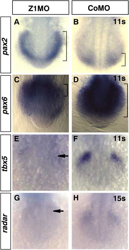
Figure 6.
Zic1 morphants display a ventralized optic vesicle. (A–H) Dorsal view, rostral/ventral to the bottom, 11s–15s. (A–B) Zic1 morphants show ectopic pax2 expression in prospective retinal tissue (brackets indicate dorsal expansion). (C,D) Pax6 retinal expression, in contrast, retracts to most dorsal retinal domains (brackets indicate retraction of pax6 expression). (E,F) Tbx5 expression in the dorsal retina is strongly reduced (arrow). (G,H) Radar expression in the dorsal retina is strongly reduced (arrow). (s) Somite stage.

