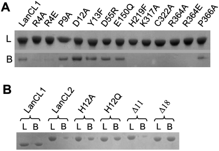
Figure 2.
Binding of LanCL1/2 variants with GSH. (A) LanCL1–GSH binding. An excessive amount (5 mg) of the protein sample was loaded onto a GSH–Sepharose4B column (0.5 mL bed volume), washed with 30 mL of PBS buffer, and optionally eluted by 1 mM GSH in 10 μM ZnCl2, 200 mM NaCl, and 20 mM Tris-HCl (pH 8.0). Lanes labeled with L (loaded) denote preincubated protein samples (∼4 μg), and those labeled with B (bound) indicate LanCL1 protein that bound to the GSH-Sepharose (from 7.5 μL of the resin slurry). Samples were analyzed by (12%) SDS-PAGE. The assay shown in the figure is representative of repeated experiments. (B) LanCL2–GSH binding and effects of the N terminus. Sample treatment was similar to A. LanCL1 was used as a positive control, followed by LanCL2 (full-length without an N-terminal tag) and its variants of point mutations and N-terminal truncations.

