Figure 3.
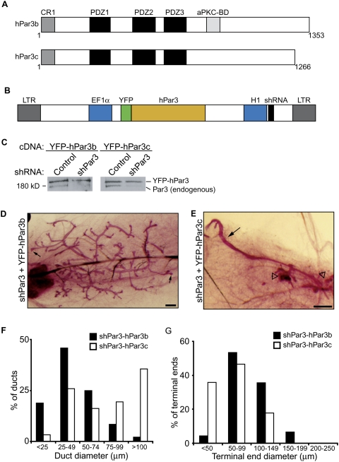
Mammary development requires aPKC-binding domain of Par3. (A) Schematic representation of human Par3b and human Par3c. Domains shown are the conserved region (CR), PSD95/Dlg/ZO1 domain (PDZ), and atypical protein kinase C-binding domain (aPKC-BD). Numbers indicate amino acid number. (B) Schematic representation of bicistronic lentiviral plasmid for expression of shRNA and cDNA. (LTR) long terminal repeat; (EF1α) EF1α promoter; (H1) H1 RNA polymerase III promoter. (C) Immunoblots of mammary epithelial lysates from cells expressing control/YFP-hPar3b, shPar3/YFP-hPar3b, control/YFPhPar3c, and shPar3/YFPhPar3c bicistronic lentiviruses. Blots were probed using anti-Par3 antibodies. The top band is YFP-hPar3 and the bottom band is endogenous Par3. (D,E) Carmine alum-stained whole-mount images of shPar3-YFPhPar3b (D) and shPar3/YFPhPar3c (E) mammary glands after 8 wk. Small arrows show end buds in D. Open arrowheads show disorganized ducts in E. Bars, 0.5 mm. (F) Distribution of duct diameters in shPar3–hPar3b and shPar3–hPar3c mammary glands (n = 3). The diameters of ducts were measured and categorized in 25-μm increments. (G) Distribution of terminal end sizes from shPar3–hPar3b and shPar3–hPar3c mammary glands (n = 3). The diameters of terminal ends were measured and categorized in 50-μm increments.

