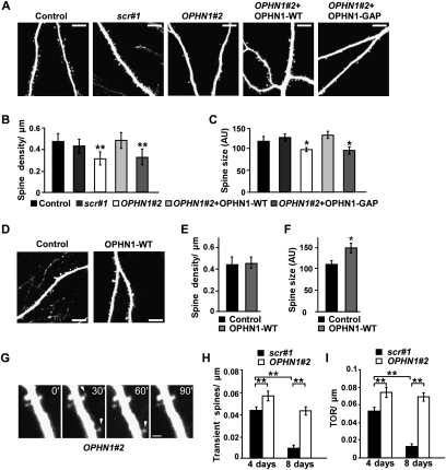
Figure 4.
OPHN1 controls maintenance of spine morphology. (A) Representative images of secondary apical dendrites from CA1 pyramidal neurons infected at 1 DIV with indicated lentiviruses, acquired with TPLSM at 8 DIV. Bars, 10 μm. (B,C) Quantification of the spine density (B) and spine size (C) for each experimental condition as indicated (control: n = 9 cells; scr#1: n = 9 cells; OPHN1#2: n = 9 cells; OPHN1#2 + OPHN1-WT: n = 8 cells; OPHN1#2 + OPHN1-GAP: n = 8 cells). (D) Representative images of secondary apical dendrites from CA1 pyramidal neurons infected at 1 DIV with lentivirus expressing EGFP only (control), or OPHN1-WT and EGFP, acquired with TPLSM at 8 DIV. Bars, 10 μm. (E,F) Quantification of spine density (E) and size (F) for control neurons and neurons expressing OPHN1-WT (n = 8 for all conditions). (G) Time-lapse images of a dendrite from a CA1 hippocampal neuron coexpressing OPHN1#2 shRNA and EGFP (at 8 d post-transfection) showing transient protrusions (arrowhead). Bar, 2 μm. (H,I) Fraction of transient spines (lifetime <1 h) (H) and spine TOR (I) in scr#1 shRNA versus OPHN1#2 shRNA-expressing neurons at 4 and 8 d post-transfection (n = 6 cells for all groups). Data are shown as mean ± SEM. (*) P < 0.05; (**) P < 0.01 by Student's t-test.

