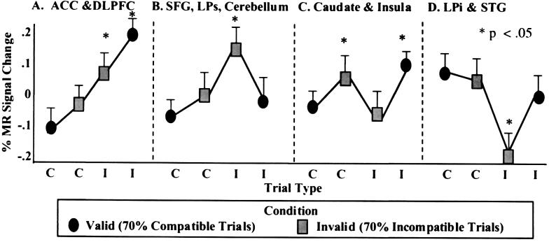
Figure 3.
Depiction of the four patterns of percentage change in normalized MR signal intensity as a function of compatible (C) and incompatible (I) trial type within the valid (70% compatible trials) and invalid (70% incompatible trials) conditions for correct trials across subjects. (A) The first pattern was observed in the anterior cingulate cortex (ACC) and dorsolateral prefrontal cortex (DLPFC). Activity in these regions increased as a function of increasing interference from the flanker stimuli and were evidenced by increasing response latencies. (B) A second pattern of results involved the right superior frontal gyrus (SFG), superior parietal lobule (LPs) and portions of the right cerebellum. These regions increased in activity during incompatible trials after consecutive incompatible trials (invalid condition). (C) A third pattern involving the basal ganglia and left insula showed increases in activity to compatible trials after consecutive incompatible trials or incompatible trials embedded in mostly compatible trials. (D) Finally, an inferior parietal (LPi) region and the superior temporal gyrus (STG) showed an inverse relation in activity to that observed in LPs and SFG. Activity in this region decreased during incompatible trials, particularly when embedded among incompatible trials (invalid condition).

