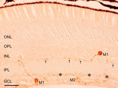
Figure 1.
A representative micrograph of melanopsin immunostained retina of adult mice. Two plexuses can be observed in the IPL, one in the innermost layer (OFF sublayer, asterisks) and the other in the outermost layer (ON sublayer, arrows). Three immunostained cells can be seen: two M1 cells (with dendritic arborization in the OFF sublayer) and one M2 cell (with dendritic arborization in the ON sublayer). GCL, ganglion cell layer; IPL, inner plexiform layer; INL, inner nuclear layer; OPL, outer plexiform layer; ONL, outer nuclear layer. Scale bar: 50 μm.

