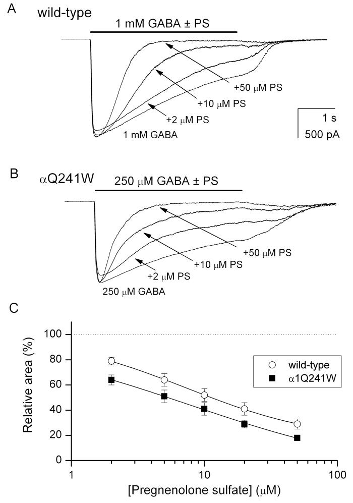
Figure 6. The α1Q241W mutation does not affect channel inhibition by pregnenolone sulfate.
(A) Sample macroscopic recordings from a cell expressing wild-type α1β2γ2L receptors. The receptors were activated by 1 mM GABA (a saturating concentration) in the absence and presence of 2, 10 or 50 μM pregnenolone sulfate (PS). The current decay phases were fitted by a single exponential to a constant level yielding 6794 ms (GABA), 4214 ms (GABA + 2 μM PS), 840 ms (GABA + 10 μM PS), and 464 ms (GABA + 50 μM PS). (B) Sample macroscopic recordings from a cell expressing α1Q241W mutant receptors. The receptors were activated by 250 μM GABA (a saturating concentration) in the absence and presence of 2, 10 or 50 μM PS. The current decay phases were fitted by a single exponential to a constant level yielding 3345 ms (GABA), 1268 ms (GABA + 2 μM PS), 864 ms (GABA + 10 μM PS), and 465 ms (GABA + 50 μM PS). (C) The relative area (total charge carried) of the macroscopic response as a function of PS concentration. The data points show mean ± SEM from 4-5 cells. The curves were fitted to: Y([steroid])=Y0 + (Ymax − Y0) [steroid]n/([steroid] + EC50)n. The best-fit parameters for the wild-type receptor were: Y0=100 % (constrained), Ymax = 15 ± 2 %, EC50 = 7.4 ± 0.4 μM, nH = 0.8 ± 0.1. The best-fit parameters for the α1Q241W mutant receptor were: Y0 = 100 % (constrained), Ymax = −12 ± 8 %, EC50 = 7.7 ± 2.2 μM, nH = 0.6 ± 0.1.

