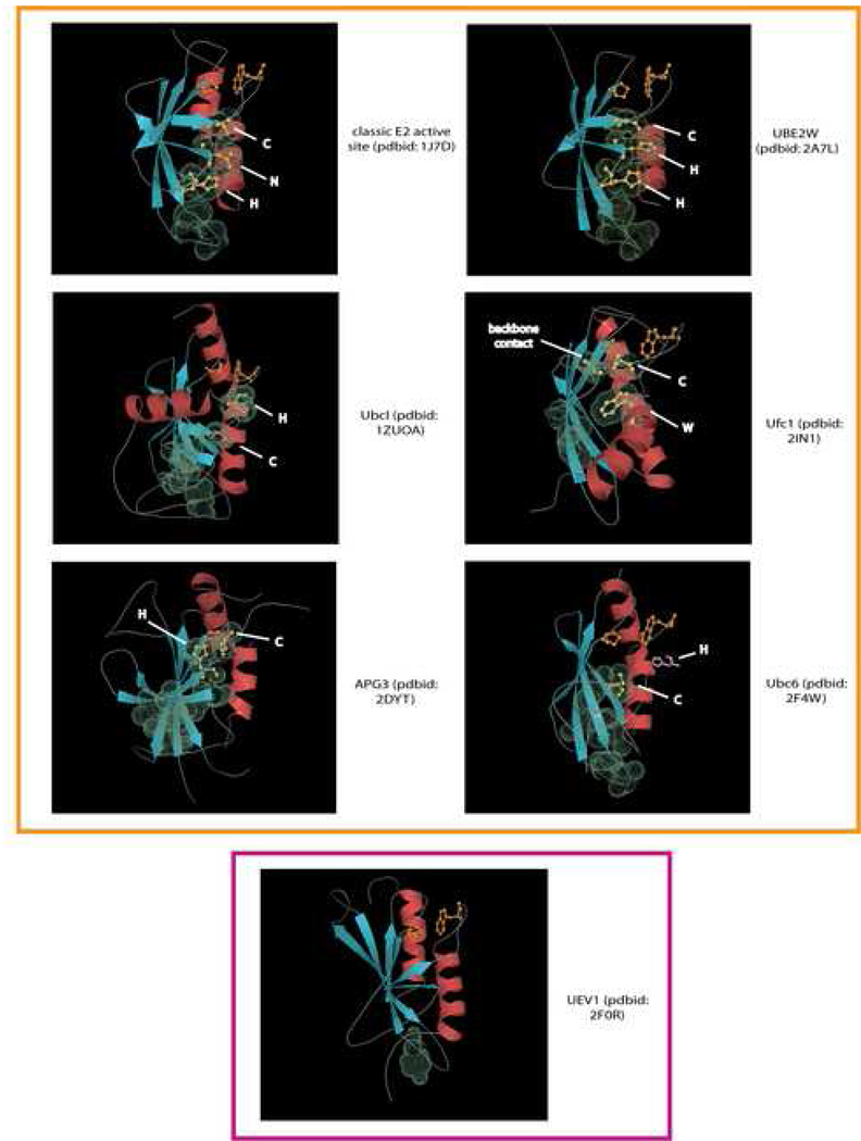
Fig. 4. Cartoon structural diagrams showing E2 domain active site variants.
Cartoon representations of X-ray structures are shown with PDB codes and family names provided at the left. Strands are colored in blue and helices in red. The catalytic cysteine and the flap asparagine/histidine or predicted equivalent residues are labeled and rendered as ball-and-sticks colored in yellow. The molecular surfaces of these residues, as well as residues predicted to form the conserved interacting chain are rendered using their van der Waal radii. The histidine residue predicted to be important in the Ubc6 family, but not observed in the crystal structure due to structure disorder is placed in its approximate predicted spatial location and colored pink. The proline and hydrophobic resides forming a conserved hydrophobic contact are rendered as ball-and-sticks and are colored orange. The active versions of the domain are boxed in orange and the inactive UEV1 family boxed in purple.

