
Fig. 4.
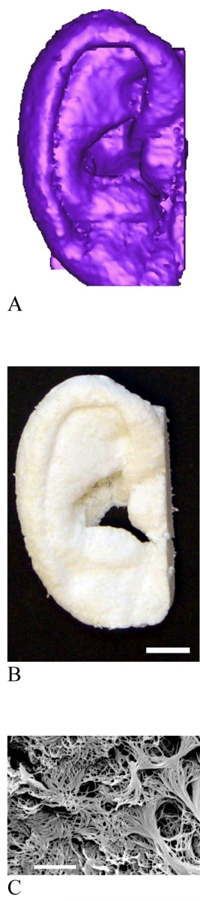
Nanofibrous scaffolds created from 3D medical images and a phase-separation technique. (A) human ear template reconstructed from histological sections; (B) resulting nanofibrous scaffold of the human ear (scale bar: 10 mm); (C) the nanofibrous pore wall morphology (scale bar: 5 μm). From Chen et al.[39] Copyright © 2006 by Elsevier. Reprinted with permission of Elsevier.