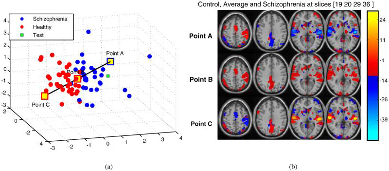
Fig. 8.

Spatial representation of the maximum separation direction,ŭ, in the reduced dimensional space. Points A-C are used to illustrate difference(s) in the activation of patient with schizophrenia, average and healthy control (from top to bottom) with 3/6 principal components at slices 19, 20, 29 and 36 (right to left) among the 46. Point A represents schizophrenia, Point B represents average, and Point C represents Healthy Control. a 3D distribution. b Regenerated slices
