Fig. 1.
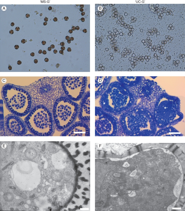
Aberrant stage of anthers and microspores of radish from (A, C, E) a sterile line, ‘MS-G’, and (B, D, F) a fertile line, ‘UC-G’. (A, B) Light micrographs of ‘UC-G’ and ‘MS-G’ at the tetrad stage in 2-mm buds. Microspores of ‘MS-G’ do not colorize compared to ‘UC-G’. (C, D) Light micrographs of toluidine blue-stained sections. The images are cross-sections of anthers in 2-mm buds. The shape of microspores is similar between ‘UC-G’ and ‘MS-G’; however, the tapetum is abnormal in ‘MS-G’ due to the presence of large vacuoles and has proliferated to fill the anther locule (*). (E, F) Transmission electron micrographs of microspores in 3-mm buds. Free microspores in ‘UC-G’ have developed sporopollenin material in their exines. In ‘MS-G’, the exines are abnormal due to irregular development and are partly empty. The cytosol of ‘MS-G’ shows plasmolysis. Scale bars: (C,D) = 50 µm; (E, F) = 500 nm.

