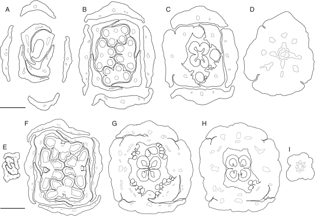
Fig. 3.
Kirkia wilmsii. Transverse microtome section series of two flower buds. Morphological surfaces drawn with thick continuous lines; secondary morphological surfaces drawn with thick dashed lines; vascular bundles drawn with thin continuous lines. (A–D) Male flower bud: (A) open sepal aestivation and imbricate petal tips; (B) contiguous (valvate) sepal aestivation and imbricate petal bases, showing two pairs of antesepalous stamens and introrse anthers with a broad and thick connective; (C) valvate sepal bases and floral cup formed by fusion of the central part of sepal bases, and petal and stamen bases, showing secretory hairs on the inner side of the petal bases and four antepetalous (delayed) sterile carpels; (D) floral base. (E–I) Sterile flower bud: (E) imbricate petal tips, arranged in pairs; (F) sepals and petals arranged in pairs, two pairs of antesepalous sterile stamens, with anthers dorsifixed basally and filament attachment hidden in a pseudopit (for term see Endress and Stumpf, 1991); (G) overlapping free sepal margins and floral cup formed by fusion of the central part of sepal bases, and petal and stamen bases, showing the carpet of secretory hairs on the inner side of the petal bases and four antepetalous carpels; (H) dorsal side of petal bases expanding between the free sepal bases and floral cup surrounding a (sterile) syncarpous and synascidiate ovary with four aborted ovules; (I) pedicel. Scale bars: A–D = 200 µm; E–I = 500 µm.

