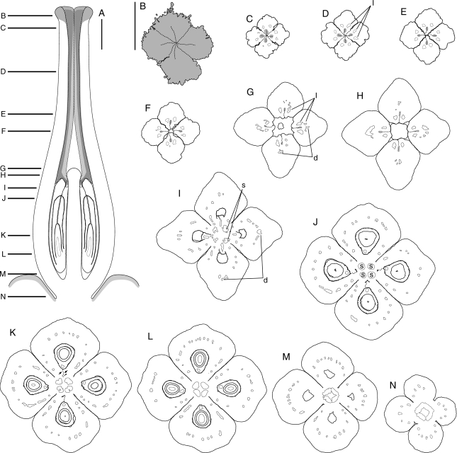
Fig. 5.
Kirkia wilmsii. Anthetic gynoecium. Morphological surfaces drawn with thick continuous lines; thick dashed lines used in (A) for parts outside the median plane of symmetry, in (B–N) for postgenitally united surfaces; vascular bundles drawn with thin continuous lines; pollen tube transmitting tract dark grey. d, Dorsal vascular bundle; l, lateral vascular bundle; s, synlateral vascular bundle. (A) Schematic median longitudinal section of gynoecium and nectary disc (light grey); postgenitally united surfaces hatched. (B–N) Transverse microtome section series; (B) stigmatic head; (C, D) postgenitally united distal parts of the carpels; (E, F) connivent but free parts of the carpels; (G, H) connivent bases of the free parts of the carpels around the hemispherical protrusion on top of the ovary; (I–M) synascidiate ovary, the two arrows in (K) pointing to the S-shaped line formed by the two lateral placentae (compare with Figs 6F and 7A); (N) gynophore. Scale bars: A, B–N = 500 µm.

