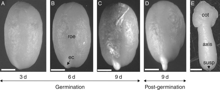
Fig. 2.
Germination and radicle protrusion of the coffee seed. (A) Seed imbibed for 3 d on water. (B) A protuberance is visible from 6 d of imbibition onwards, showing the endosperm cap (ec) and rest of the endosperm (roe). (C) A more prominent protuberance just before radicle protrusion. (D) Radicle protrusion starts after 5 d of imbibition on water, and following germination the radicle grows and the endosperm remains attached to the cotyledons. (E) Imbibed coffee embryo isolated after 6 d showing the cotyledons (cot), the embryonic axis and remnants of the suspensor (susp) on the radicle tip. Sacle bars: (A–D) = 2 mm; (E) = 0·5 mm.

