
Fig. 3.
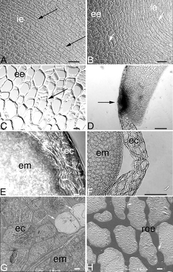
Light micrographs of the endosperm cap and rest of the endosperm of coffee seed during germination. (A) Cells of the rest of the endosperm adjacent to the embryo during imbibition. Note the high degree of uniformity of cell size in this region (arrows); ie, internal endosperm. (B) Cells of the rest of the endosperm during imbibition showing differences in cell morphology in internal (ie) and external (ee) endosperm. The cells of the internal endosperm (arrow) adjacent to the embryo are rectangular in shape and will be the first cells to be consumed following germination. The cells of the external endosperm have a polygonal shape (arrow); these cells will be consumed later. (C) Higher magnification of the external endosperm region of the rest of the endosperm, showing thin-walled areas (arrow). (D) Endosperm cap of a 9-d imbibed seed showing remnants of a suspensor at the radicle tip (dark spot, arrowed). (E) Endosperm cap (ec) region and embryo (em) after 6 d of imbibition, showing compressed cells in the endosperm. (F) Endosperm cap (ec) region and embryo (em) in a 9-d imbibed seed, showing loss of cell integrity just before radicle protrusion. (G) Endosperm cap of a 6-d imbibed seed showing thinner cell walls. Note that some cells are not completely hydrated (arrows) and are surrounded by fully hydrated cells of the endosperm cap (ec) and embryo (em). (H) Cells of the rest of the endosperm (roe) of 6-d imbibed seeds. Note that these cell walls are thicker than the cell walls of the endosperm cap region shown in (G). Scale bars: (C, E, G) = 10 µm; (A, B, D, F, H) = 100 µm.