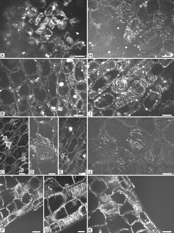
Fig. 7.

Fluorescence micrographs of longitudinal sections of embryos from water- and ABA-imbibed seed during and following germination. (A) Axis region of dry embryo (0 d of imbibition). Dots represent clusters of preserved β-tubulin. (B) Embryonic axis region from 3-d imbibed seeds on water, showing low microtubule abundance and random microtubular organization. (C–E) Embryonic axis regions from seeds imbibed on water for 6 d, showing transversal microtubular orientation in the outer cortex cells, and mitotic figures before radicle protrusion. (F) Embryonic axis region from 9-d water-imbibed seeds showing transversal microtubular orientation in the epidermal cells. (G) Embryonic axis region after radicle protrusion, showing transversal microtubular orientation and higher abundance of microtubules. (H) Embryonic axis region from 3-d imbibed seeds on ABA showing low abundance of microtubules. (I) Embryonic axis region from 6-d ABA-imbibed seeds, showing random orientation of the microtubules. (J) Embryonic axis region from 9-d imbibed seeds on ABA showing low abundance of microtubules. (K) Embryonic axis region from 9-d ABA-imbibed seeds that were rinsed in demineralised water and transferred to water for 30 d. Note that the microtubules reappeared as in water-imbibed seeds. Scale bars = 2 µm.
