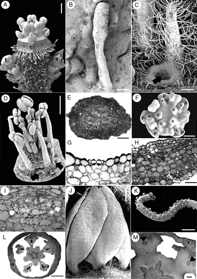
Fig. 6.

Suriana maritima flower and inflorescence morphology. A–D, F, J and K are SEM micrographs; E, G–I, L and M are transverse sections. (A) Lateral view of a dichasial cyme; the terminal flower is surrounded by two partial inflorescences, each repeating the dichasial cyme structure. (B) Glandular multicellular hair growing on sepal. (C) Dorsal view of a mature hairy staminode with extended ring-shaped tissue surrounding petal base (black arrow, petal detached). (D) Lateral view of mature androecium and gynoecium; thickened petal bases with and without associated staminodes (black arrows). (E) Pedicel. (F) Polar view of floral bud with missing carpel and four staminodes (asterisks). (G) Section of a sepal showing a single stomata in the abaxial (ventral) epidermis. (H) Sepal; ventral side towards the upper side of the figure. (I) Sepal vascular bundle surrounded by tissue with oxalate druses. (J) Lateral view of preanthetic bud with contorted petal aestivation. (K) Ornamented hair from petal. (L) Floral receptacle with petal and staminodial tissue differentiating. Petals (their vascular bundles indicated by arrows) surrounded by the base of the staminodes (white asterisks). Stamens are indicated by black asterisks. (M) Petal bases surrounded by staminode base; arrowheads indicate petal vascular tissue. c, carpel; p, petal; r, receptacle; s, sepal; sy, style; t, terminal flower. Scale bars: A, C = 200 µm; B, G = 20 µm; D, L = 1 mm; E, M =100 µm; F = 150 µm; H = 50 µm; I = 2 µm; J = 0·5 mm; K = 10 µm.
