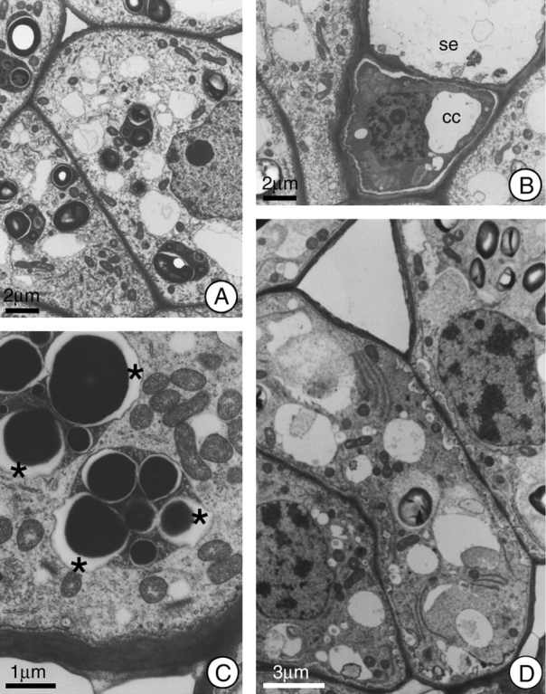
Fig. 2.

General view of the cells in the secretory region of the nectary of H. stigonocarpa. (A) Secretory cell 2 h before anthesis; note dense cytoplasm, presence of mitochondria, small vacuoles, and plastids with well developed starch grains and dense stroma. (B) Sieve element (se) and companion cell (cc); note absence of symplastic connections between these cells and the secretory cells adjacent to them. (C) Secretory cell at anthesis; note the elevated number of mitochondria and the formation of lacuna (*) next to the starch grains within the plastids. (D) Secretory cell 2 h after anthesis; note the incorporation of the plastids into the cytoplasmic matrix and the increase in periplasmic space. Scale bars: 2 µm (A and B); 1 µm (C); 3 µm (D).
