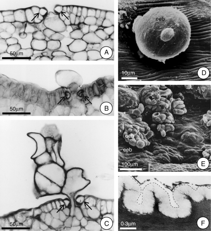
Fig. 5.

Extrastomatic bodies (eeb) formed after the secretory phase, and detail of the epidermal cell showing the cuticle. (A–C) Proposed sequence of the formation of the extrastomatic bodies (arrows indicate guard cells). Note in A the initial expansion of the cell towards the stomatal pore. (D) Young extrastomatic body, under SEM. (E) Fully differentiated extrastomatic bodies, under SEM. (F) Detail (under TEM) of the epidermal cell showing the cuticle; the dotted line indicates the cuticle outline. Scale bars: 50 µm (A–C); 10 µm (D); 100 µm (E); 0·3 µm (F).
